Author: Alessio Liberati
As already seen in the previous clinical case, ankle sprain is a subluxative phenomenon of the tibio-tarsal joint during which the capsular-ligament compartment is heavily stressed.
Ankle sprain (AS) is a very common injury among athletes and the general population. In the United States, for example, it has an incidence rate of 2.15 per 1,000 people per year. Almost half of all AS (49.3%) occur during athletic activity: basketball 41.1%, American football 9.3% and soccer 7.9%, have the highest injury rates [1].
There is strong evidence that, after an initial ankle sprain, the recurrence rate doubles, especially during the first year after injury [2].
Anamnesis
A 30-year-old man presented an inversion sprain occurred while running, on a left ankle that had already been injured 2 years ago. The traumatic event took place 7 days before the day of the examination: since then the patient reported pain on loading (VAS 4/10) with a feeling of instability and ankle that "gives way" with consequent limitation in walking. When unloading, the patient had no pain, but the feeling of instability remained.
Evaluation
On physical examination the ankle presented:
- pain on palpation of the anterior peroneal astragalic ligament and to a lesser extent also the deltoid ligament
- absence of edema
- limitation of passive range of motion (ROM) in plantar flexion and supination associated with pain
In addition, the patient performed the following tests with baiobit support:
1) MONOPODAL BALANCE TEST
It was used to assess the proprioceptive ability of the healthy limb compared to the injured limb. The different results, between the right and left lower limb, suggested a proprioception deficit at the level of the injured ankle.
| Monopodal right | Monopodal left |
Area of ellipse [mm2]
| 637
| 13.161
|
AP Oscillation [mm]
| 40
| 114
|
ML Oscillation [mm]
| 18
| 17 |
2) WALKING TEST
The test revealed the subject's difficulty in equally dividing the various phases of the step, with a reduced support phase of the injured lower limb, equal to 48.22% compared to the expected normal value of 60%. This deficit was most probably due to an altered propulsion of the injured limb equal to 7.5 which was lower than the contralateral limb, showing a tendency to avoid loading the injured ankle. The gait symmetry index of 38.4%, compared with an expected value of more than 90%, showed an important difference between the right and left gait cycles.
3) MONOPODAL COUNTER MOVEMENT JUMP TEST
The aim was to evaluate the performance of the injured AI compared to the healthy limb’s one during high-load activity. This test showed the subject's difficulty in developing strength and power with the injured limb. In addition, the test detected the lack of reactivity and elasticity of the injured side.
| Monopodal left | Monopodal right |
Height [cm]
| 4
| 19
|
Force [KN]
| 0,7
| 1,22
|
Power [W]
| 984,57
| 1.625,19
|
Reactivity index | 0,19
| 0,34 |
Rehabilitation program
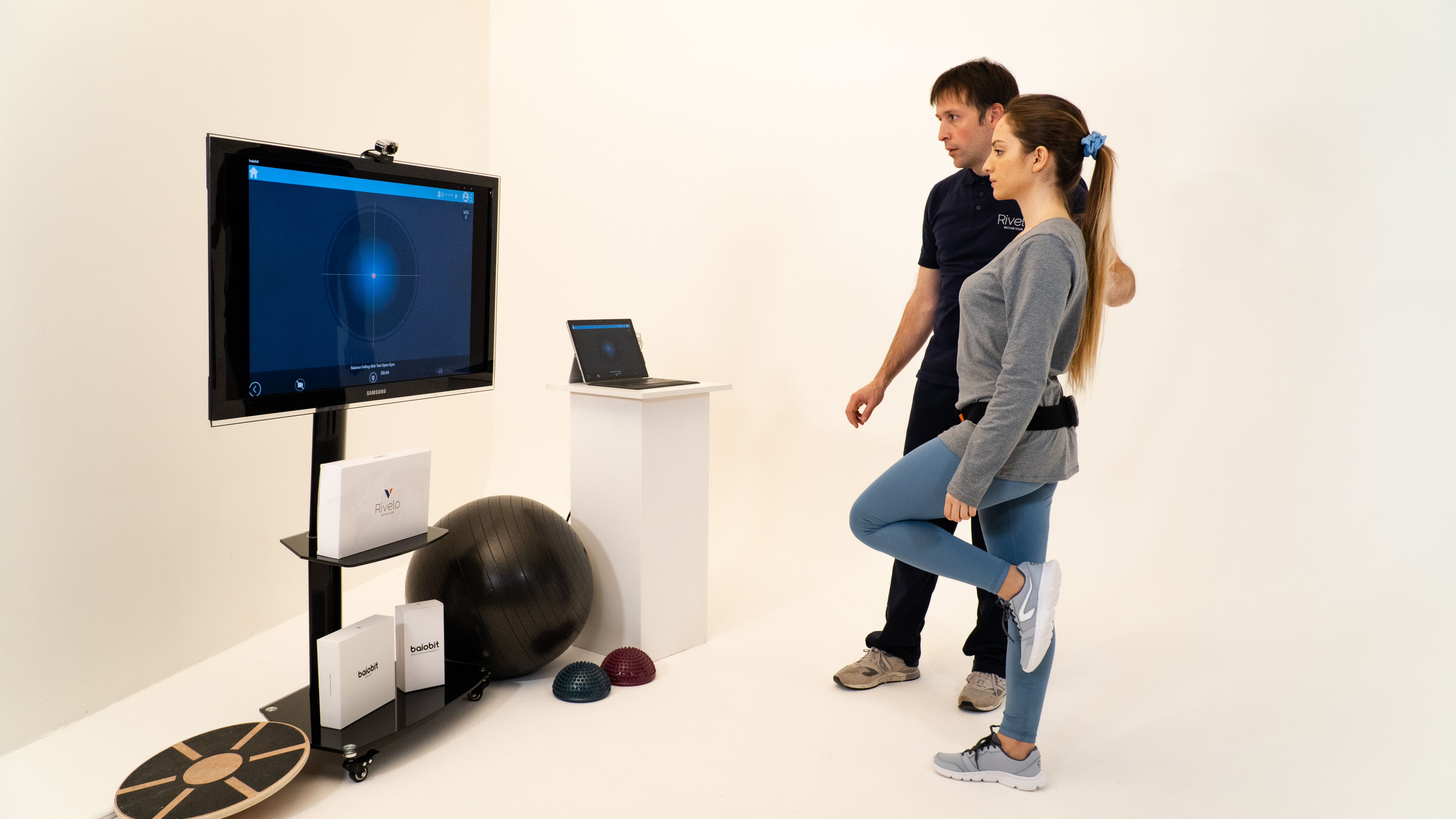
A rehabilitation program of 3 weekly sessions was set up, consisting of manual treatment with Fascial Manipulation and therapeutic exercises to strengthen muscles and improve proprioception. To promote proprioceptive recovery [3] and the correct maintenance of balance, static monopodal balance exercises were included. These tests were performed first with eyes open and then with eyes closed. The patient was guided by biofeedback to controlled displacements of his centre of mass (exercise is shown in the following image - Fig. 1).

Fig. 1: Example of biofeedback balance exercise
Results
At the end of the 3 rehabilitation sessions, a re-evaluation of the outcomes was carried out repeating the initial tests performed with baibobit:
1) MONOPODAL BALANCE TEST
This allowed to evaluate the recovery evolution of the injured limb before and after treatment comparing the results with the values of the healthy.
| Monopodal right | Monopodal left pre-treatment | Monopodal left post treatment |
Area of ellipse [mm2]
| 637
| 13.161
| 1.117
|
AP oscillation [mm]
| 40
| 114
| 53
|
ML oscillation [mm]
| 18
| 178
| 22 |
The results showed an improvement in the injured limb before and after treatment, but a marked difference between the right and left limb. For this reason, it was advisability continuing the rehabilitation to improve the proprioceptive aspect.
2) WALKING TEST
Compared to the initial test, there was an increase in symmetry and a normalization of the left AI stance phase:
- symmetry 96.7%
- left support phase 60.43%
3) MONOPODAL COUNTER MOVEMENT JUMP TEST
There was a realignment between the right and left AI values:
| Monopodal left | Monopodal right |
Height [cm]
| 19
| 21
|
Force [KN]
| 1,35
| 1,35
|
Power [W]
| 1.686,9
| 1.787,7
|
Reactivity index | 0,29
| 0,28 |
The measurements showed a clear improvement in the injured lower limb. The slight difference from the healthy limb is lower than 10%, so the improvement is acceptable.
Bibliography
[1] Waterman, B. R., Owens, B. D., Davey, S., Zacchilli, M. A., & Belmont Jr, P. J. (2010). The epidemiology of ankle sprains in the United States. JBJS, 92(13), 2279-2284.
[2] Trevino, S. G., Davis, P., & Hecht, P. J. (1994). Management of acute and chronic lateral ligament injuries of the ankle. Orthopedic Clinics of North America, 25(1), 1-16.
[3] Verhagen, E., Van Der Beek, A., Twisk, J., Bouter, L., Bahr, R., & Van Mechelen, W. (2004). The effect of a proprioceptive balance board training program for the prevention of ankle sprains: a prospective controlled trial. The American journal of sports medicine, 32(6), 1385-1393.